Written by the Core Power Peptides Research Team — biochemists and laboratory specialists compiling and verifying peptide research data. Learn about our quality standards →
Myostatin — formally designated Growth Differentiation Factor 8 (GDF-8) — is a member of the transforming growth factor-beta (TGF-β) superfamily that functions as the primary endogenous inhibitor of skeletal muscle growth. Discovered by McPherron et al. in 1997, myostatin has become one of the most studied proteins in muscle biology due to the dramatic phenotype of animals and humans with loss-of-function mutations: uniform, severe muscle hypertrophy with no other apparent pathology. In research settings, recombinant myostatin protein is used as a tool to study skeletal muscle regulation, muscle wasting diseases, and the myostatin-follistatin signaling axis.
Search interest in “myostatin peptides” has climbed steadily throughout 2025–2026, reflecting both academic expansion and the growing market for tools to investigate muscle physiology at the molecular level.
What Is Myostatin? Biology and Function
Myostatin is a secreted homodimeric protein that signals through a defined receptor pathway to suppress muscle growth:
- Gene: MSTN (chromosome 2q32.2 in humans)
- Expression: Primarily skeletal muscle; lower levels in cardiac muscle, adipose tissue
- Secretion: Produced by myocytes and secreted into circulation as a latent complex; proteolytically activated by BMP-1 and related tolloid-like proteases
- Circulating form: Latent myostatin propeptide complex (inactive) and small amounts of active mature myostatin dimer
Mechanism of Action
Myostatin signals through the Activin receptor type IIB (ActRIIB) → ALK4/ALK5 → Smad2/3 pathway:
- ActRIIB Binding: Mature myostatin dimer binds ActRIIB with high affinity (Kd ~1 nM), recruiting the type I receptor (ALK4 or ALK5)
- Smad2/3 Phosphorylation: The type I receptor phosphorylates Smad2 and Smad3, which translocate to the nucleus and activate transcription of muscle atrophy genes
- MuRF1 and MAFbx/Atrogin-1: Key E3 ubiquitin ligases upregulated by Smad2/3 signaling — these drive proteasomal degradation of myofibrillar proteins (actin, myosin), producing muscle atrophy
- mTOR/AKT Suppression: Myostatin signaling inhibits AKT/mTOR, the primary pro-anabolic pathway regulating protein synthesis and satellite cell activation
- Satellite Cell Inhibition: Myostatin inhibits the self-renewal and proliferation of muscle satellite cells (stem cells), reducing regenerative capacity
- Adipogenesis Promotion: In adipose tissue, myostatin promotes preadipocyte differentiation and fat accumulation — explaining the lean phenotype of myostatin-null animals beyond just muscle hypertrophy
Myostatin Loss-of-Function: Natural Evidence
The clearest demonstration of myostatin’s role comes from naturally occurring mutations:
- Belgian Blue and Piedmontese cattle: Natural myostatin mutations produce “double-muscling” — 20–40% more muscle mass than unaffected animals, with virtually no fat deposition
- Whippet dogs (bully whippet): Homozygous myostatin deletion produces extreme muscularity; heterozygotes are elite racing dogs with enhanced muscle performance
- Human case (2004, NEJM): An infant with bilateral myostatin loss-of-function mutation demonstrated extraordinary muscle development with no apparent adverse effects at 4.5 years
Research Applications for Recombinant Myostatin
Recombinant myostatin protein is used as a research reagent to establish the myostatin axis in new model systems and as a comparator in inhibitor screening:
Muscle Atrophy and Wasting Disease Models
- Establishing in vitro muscle atrophy: C2C12 myotube treatment with recombinant myostatin produces dose-dependent atrophy, validated by decreased myotube diameter and upregulation of atrogenes (MuRF1, MAFbx)
- Cachexia research: Myostatin is elevated in cancer cachexia and is used as a target biomarker in tumor-muscle axis studies
- Sarcopenia models: Circulating myostatin increases with aging; recombinant myostatin is used to induce accelerated sarcopenia phenotypes in young rodent and cell models
- DMD/muscular dystrophy: Myostatin inhibition is a therapeutic target in Duchenne muscular dystrophy research
Myostatin Inhibitor Screening
- Follistatin: The primary endogenous myostatin antagonist. Studies quantifying myostatin/follistatin balance use recombinant myostatin as the positive control challenge
- Anti-myostatin antibody research: Multiple biologics targeting myostatin (domagrozumab, landogrozumab, trevogrumab) have entered clinical trials — preclinical work uses recombinant myostatin to validate blocking activity
- Small molecule ActRIIB inhibitors: Drug discovery programs use recombinant myostatin in cell-based assays to screen compounds for Smad2/3 pathway inhibition
Adipose-Muscle Crosstalk
- Myostatin’s dual role in muscle and adipose regulation makes it a key reagent for studying the metabolic intersection of muscle wasting and fat accumulation
- Preadipocyte differentiation assays: Recombinant myostatin accelerates 3T3-L1 adipogenesis in a dose-dependent manner
Key Published Research
- McPherron et al. (Nature, 1997): Discovery of myostatin — MSTN knockout mice have 2–3× normal muscle mass
- Zimmers et al. (Science, 2002): Systemic myostatin overexpression produces profound muscle wasting, establishing myostatin as a cachexia mediator
- Lee & McPherron (PNAS, 2001): Follistatin overexpression doubles muscle mass in mice — more than myostatin knockout alone — establishing follistatin/myostatin balance as the key regulatory axis
- Trendelenburg et al. (Skeletal Muscle, 2012): Comprehensive characterization of myostatin signaling in human primary myotubes at physiologically relevant concentrations
Dosage Reference for Research
| Research Context | Dose | Route | Notes |
|---|
| C2C12 atrophy (in vitro) | 10–200 ng/mL | Culture media | Dose-dependent atrophy induction |
| Primary human myotubes | 50–500 ng/mL | Culture media | Smad2/3 phosphorylation assays |
| Adipogenesis (3T3-L1) | 10–100 ng/mL | Differentiation media | Lipid accumulation assays |
| In vivo (systemic wasting, rodent) | 1–5 µg/kg/day | SC or IP | Cachexia model induction |
Myostatin Inhibition vs. Myostatin Supplementation — Why Use Recombinant Protein?
Most commercial interest is in inhibiting myostatin. So why use recombinant myostatin protein in research?
- Positive control: Any myostatin inhibitor study needs a validated myostatin challenge to test against. Recombinant myostatin establishes the baseline atrophy or signaling state that the inhibitor reverses
- Pathway validation: Before testing a novel inhibitor, researchers confirm the signaling pathway (Smad2/3, mTOR suppression) using recombinant protein
- Dose-response modeling: Understanding myostatin concentration-response relationships in a specific cell type or model system is prerequisite to inhibitor dose selection
- Biomarker research: Circulating myostatin levels are a biomarker of muscle health. Assay calibration requires high-purity recombinant standard
Where to Buy Myostatin (GDF-8) for Research
Recombinant myostatin is a larger protein than typical synthetic peptides, requiring more stringent quality verification:
- SDS-PAGE or SEC-HPLC purity documentation (target: ≥95%+ for a 25kDa recombinant protein)
- Biological activity validation: manufacturer-supplied ED50 data from Smad2/3 phosphorylation or muscle atrophy assay
- Endotoxin testing critical for cell-based assays (LPS contamination from E. coli expression systems can confound inflammation measurements)
- Proper storage: recombinant proteins require −80°C storage for long-term stability; avoid freeze-thaw cycles
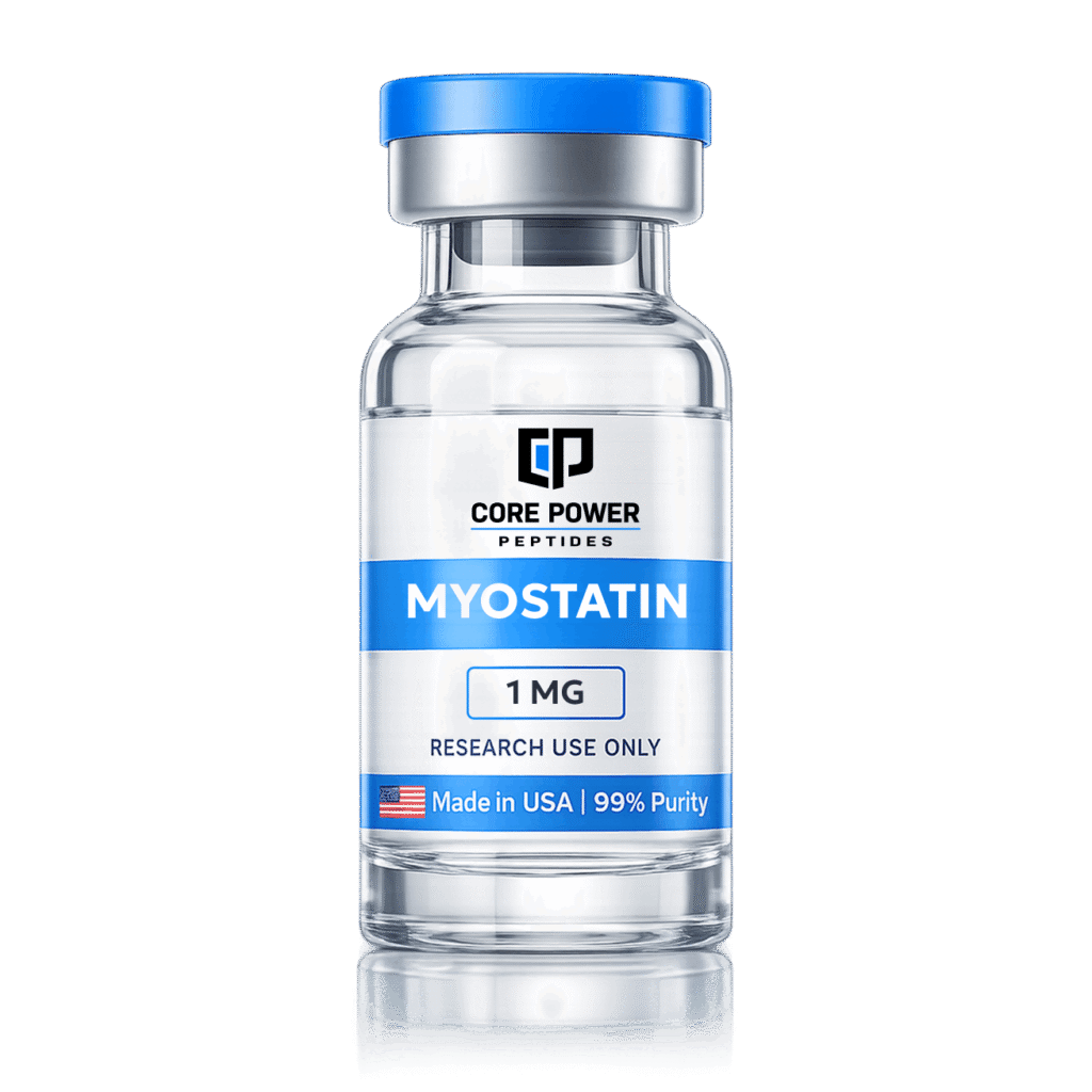
Myostatin (GDF-8) 1mg
Purity: 95%+ SDS-PAGE
Format: Lyophilized Powder
Testing: Third-Party COA
$85
Buy Now
Related Research Peptides
Researchers studying the myostatin signaling axis typically work with these complementary compounds:
HPLC VerifiedFollistatin 344 — 1mg
Purity: 95%+ HPLC
Role: Primary myostatin antagonist
Testing: Third-Party COA
$280
Buy Now
HPLC VerifiedAOD 9604 10mg
Purity: 99%+ HPLC
Role: Lipolysis / body composition
Testing: Third-Party COA
$165
Buy Now
Frequently Asked Questions
Is myostatin a peptide?
Myostatin (GDF-8) is a protein, not a short peptide. At ~25 kDa in its mature dimeric form, it is significantly larger than typical research peptides (which are usually under 10 kDa). Recombinant myostatin for research use is expressed in E. coli or mammalian cell systems and purified to research grade, not chemically synthesized like most peptides.
What is the opposite of myostatin?
Follistatin is the primary endogenous antagonist of myostatin. It binds and neutralizes myostatin (and activin) with high affinity, preventing ActRIIB receptor engagement. The balance between circulating myostatin and follistatin is a critical determinant of muscle mass. Exercise increases follistatin levels and decreases myostatin — this is one mechanism by which resistance training promotes hypertrophy.
Does myostatin research apply to humans?
The myostatin pathway is highly conserved across mammals and appears functionally identical in humans. Multiple clinical trials have investigated myostatin inhibition in sarcopenia, cancer cachexia, and muscular dystrophy — validating the pathway’s relevance. However, no myostatin-targeting therapy has achieved FDA approval as of 2026.
Scientific reference only. Recombinant myostatin from Core Power Peptides is supplied for in vitro and laboratory research — not for human consumption, and not a substitute for medical guidance. Researchers remain responsible for applicable local regulations.